What is Competitive ELISA
Competitive ELISA (also known as inhibition or blocking ELISA) measures the concentration of an analyte in a sample by quantifying its interference with an expected signal. This method is performed by coating a microplate with either an analyte or an antibody specific to the target analyte (direct and indirect methods). Then, a partially detected target analyte is added to compete for binding sites on the antibody. The signal is inversely proportional to the analyte concentration; thus, when the target analyte concentration is high, it competitively binds to a limited amount of labeled antibody, resulting in a weaker reference signal compared to a lower analyte concentration.
The theoretical basis of competitive ELISA is the use of a limited amount of antibody, which creates a competitive relationship between two antigens. This method is commonly used for detecting small molecules with fewer epitopes.
Experimental Overview
Taking direct competitive ELISA as an example:
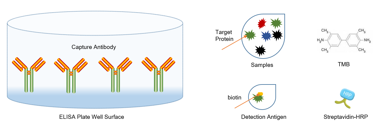
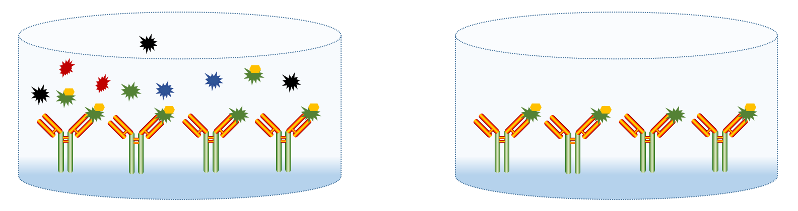
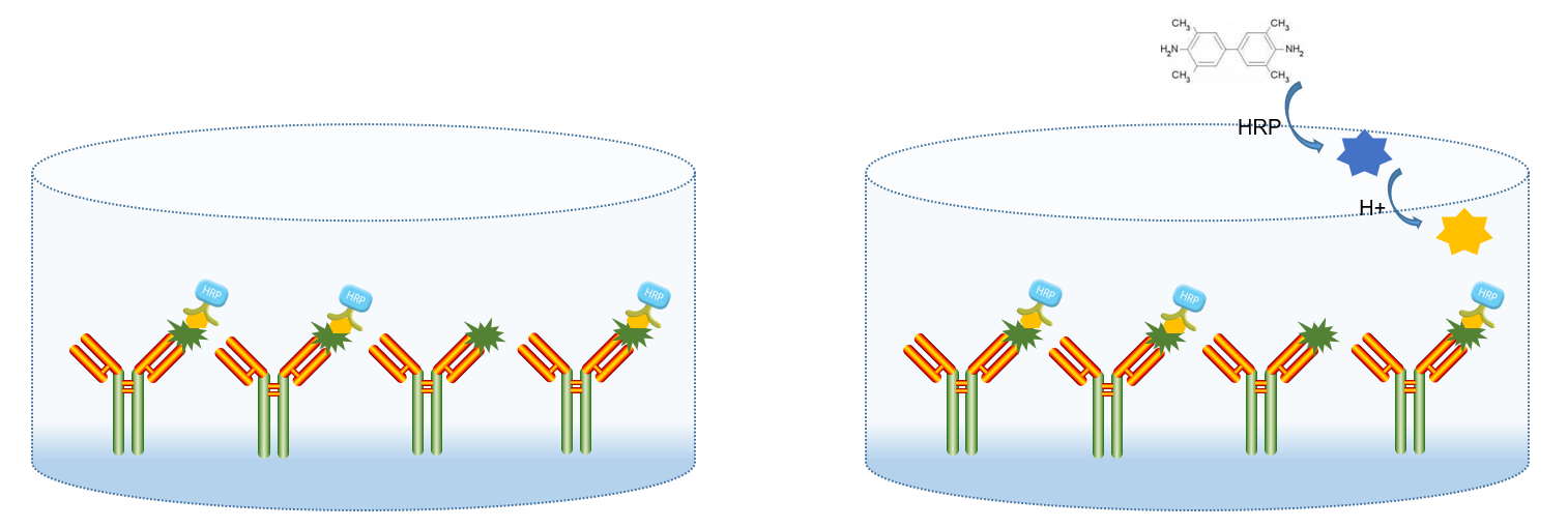
A specific antibody is adsorbed onto a solid-phase carrier, and a sample (standard or unknown) along with a biotin-labeled detection antigen is added. These two components compete to bind with the solid-phase antibody. After incubation, horseradish peroxidase (HRP)-labeled streptavidin is added, followed by thorough washing and the addition of a colorimetric substrate. The intensity of the developed color is inversely proportional to the concentration of the analyte in the sample.
Steps
- Prepare all reagents and prepare gradient-diluted standards. Add washing solution to the strips and let them soak for 30 seconds.
- Add 2-fold serially diluted standards to the standard wells. Add standard dilution buffer or culture medium to the non-specific binding (NSB) and maximum binding (B0) wells.
- Serum/plasma: Add samples to the sample wells. Cell culture supernatant: Add culture supernatant to the sample wells.
- Except for Blank and NSB wells, add 1× detection buffer to NSB wells and diluted conjugate to all other wells.
- Cover the plate, incubate at room temperature for 1 hour, and wash 6 times.
- Except for TA and Blank wells, add diluted HRP-labeled streptavidin to the remaining wells.
- Cover the plate, incubate at room temperature for 30 minutes, and wash 6 times.
- Add diluted HRP-labeled streptavidin to the TA wells.
- Add colorimetric substrate to each well, incubate at room temperature in the dark for 5-30 minutes.
- Add stop solution to each well.
- Measure the optical density (OD) at 450 nm within 30 minutes, with a reference wavelength of 570 nm or 630 nm.
Note: The above steps are based on Lianke Bio’s ELISA kit. Different manufacturers may have different procedures, so always read the product manual carefully before starting the experiment.
Differences Between Direct and Indirect Competitive ELISA
Labeling
- Direct Competitive ELISA: Labeled antigen competes with the antigen in the sample for antibody binding.
- Indirect Competitive ELISA: Labeled antibody competes with the antigen in the sample for binding to the solid-phase antigen.
Model
- Direct Competitive ELISA: The plate is coated with an antibody. HRP-labeled antigen (HRP-Ag) and the sample are added together. The antigen in the sample competes with HRP-Ag for binding to the coated antibody. The amount of HRP-Ag bound to the solid phase is inversely proportional to the antigen concentration in the sample.
- Indirect Competitive ELISA: The plate is coated with an antigen. HRP-labeled antibody (HRP-Ab) and the sample are added together. The antigen in the sample competes with the coated antigen for binding to HRP-Ab. The amount of HRP-Ab bound to the solid phase is inversely proportional to the antigen concentration in the sample.
Binding Opportunities
- Direct Competitive ELISA: Both labeled and target antigens are in the liquid phase, so they have equal binding opportunities with the antibody.
- Indirect Competitive ELISA: The coated antigen has a smaller contact area with the antibody. Binding opportunities between the coated antigen and the antibody are unequal, following a sequential saturation model—only the remaining antibodies unbound to the target antigen will bind to the coated antigen.
Signal Amplification
- Direct Competitive ELISA: Generally difficult to amplify; commonly used biotinylated antigens with enzyme-labeled streptavidin.
- Indirect Competitive ELISA: Can use enzyme-labeled secondary antibodies for signal amplification.
Sensitivity
- Indirect Competitive ELISA is more sensitive than Direct Competitive ELISA.
- Due to differences in binding opportunities, the inhibition rate of the indirect method is greater than that of the direct method, resulting in a steeper inhibition curve and higher sensitivity. Indirect ELISA can further enhance sensitivity by using enzyme-labeled secondary antibodies for amplification.
Additional Notes
Competitive ELISA can also be used to detect large molecule antigens or even antibodies.
- When detecting large molecule antigens, spatial hindrance affects detection sensitivity, making this method less sensitive than the sandwich ELISA.
- When detecting antibodies, the competitive relationship between two antibodies may not be ideal, leading to lower reliability of results.