2 - ELISA Weak/No Signal? Causes & Solutions for Reliable Results

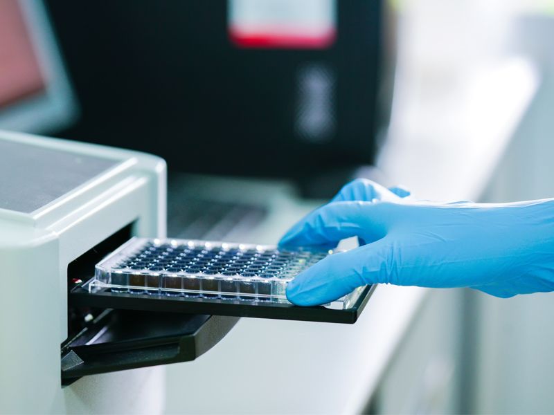
Are you getting no signal or weak signals in your ELISA assays? This common problem can derail your research. Discover the key causes and proven solutions to restore your assay sensitivity: Top 10 Reasons for ELISA Signal Failure & How to Fix Them:

  • Mixed reagent batches
  • Improper storage
  • Expired reagents
  • Missing detection antibodies
  • Incorrect incubation
  • Wrong addition order
  • Contaminated reagents
  • Pipetting errors
  • Microsphere issues
  • Inadequate washing
Critical Tip: For one-step ELISAs, control humidity exposure and loading intervals to prevent microsphere degradation.

Continue Reading2 - ELISA Weak/No Signal? Causes & Solutions for Reliable Results

3 - ELISA Standard Curve Problems? Fix Poor Linearity & Gradient Issues

Is your ELISA standard curve showing poor linearity or no gradient distribution? This critical assay problem can compromise your data reliability. Learn the root causes and expert solutions: Top Causes & Solutions for Standard Curve Failures:

  • Incorrect incubation
  • Improper washing
  • Pipetting errors
  • Microsphere problems
  • Humidity exposure
Pro Tip: Always include a positive control to validate curve performance before sample testing. Why This Matters: A properly gradiented standard curve is essential for accurate quantification in Biomedical research, Clinical diagnostics and Drug development

Continue Reading3 - ELISA Standard Curve Problems? Fix Poor Linearity & Gradient Issues

A Brife Review of the Apoptosis Pathway

Apoptosis occurs through two main pathways: extrinsic and intrinsic. The extrinsic pathway activates caspases via extracellular signals, while the intrinsic pathway involves cytochrome c release from mitochondria, forming an apoptosome with Apaf-1 and caspase-9 to activate caspase-3. Caspases, cysteine proteases, degrade key intracellular proteins, leading to apoptosis. They are divided into the ICE subfamily (involved in inflammation) and the CED-3 family (involved in apoptosis). The mitochondrial pathway is regulated by Bcl-2 family proteins, which control the permeability transition pore (PT pore) and cytochrome c release. Additional proteins like Smac, AIF, and Endo G also contribute to apoptosis. In Fas signaling, caspase-8 activation amplifies the apoptotic signal through Bid cleavage, particularly in type II cells.

Continue ReadingA Brife Review of the Apoptosis Pathway

Tumor necrosis factor (TNF): TNF-α & TNF-β

The Tumor Necrosis Factor (TNF) family comprises cytokines that induce cell death (apoptosis). Key members include TNF-α and TNF-β. TNF-α, a pro-inflammatory cytokine, is secreted by various cells like macrophages, T cells, and adipocytes. It signals through TNFR1 and TNFR2 receptors, mediating immune responses, tumor cytotoxicity, and roles in septic shock, autoimmune diseases, and inflammation. TNF-β, produced by activated T and B cells, shares similar functions with TNF-α, including immune regulation, cell proliferation, apoptosis, and tumor cytotoxicity. Both TNF-α and TNF-β exhibit significant cross-reactivity between humans and mice, highlighting their conserved biological roles.

Continue ReadingTumor necrosis factor (TNF): TNF-α & TNF-β

【Flow cytometry】A Review of Apoptosis Data Analysis Techniques

The concept of apoptosis and its characteristics Apoptosis: also known as programmed cell death (PCD), is the autonomous and orderly death of cells controlled by genes in order to maintain the stability of the internal environment, which plays a necessary role in the removal of unwanted or abnormal cells by multicellular organisms, and belongs to the "suicide" behavior of cells. Typical features of apoptosis loss of mitochondrial membrane potential; eversion of cell membrane PS (phosphatidylserine); Nuclei condense and rupture. Apoptosis Programmed death is an active death process taken by the body to better adapt to the living environment. Heat analysis of apoptosis research Apoptosis and disease Insufficient ApoptosisExcessive ApoptosisNeoplasmsCardiovascular DiseasesAutoimmune DiseasesHematologic DiseasesViral InfectionWith Severe HepatitisDegenerative Neurological DiseasesSuch as Alzheimer's DiseaseParkinson's…

Continue Reading【Flow cytometry】A Review of Apoptosis Data Analysis Techniques

ELISA (Enzyme-Linked Immunosorbent Assay): Principle, Types, and Step-by-Step Protocol

Learn about ELISA (Enzyme-Linked Immunosorbent Assay), a highly sensitive immunoassay technique used in medical diagnostics and biopharmaceuticals. This guide covers its principles, common types (sandwich, direct, indirect, competitive), detailed protocol, and key considerations for accurate results.

Continue ReadingELISA (Enzyme-Linked Immunosorbent Assay): Principle, Types, and Step-by-Step Protocol

Flow Cytometry Cell Cycle Analysis: PI Staining, Fixation vs. Live Cell Protocols & Key Considerations

Learn the principles of cell cycle analysis by flow cytometry, including PI staining, RNase treatment, critical no-wash steps, and protocols for fixed & live cells. Includes step-by-step methods and troubleshooting tips for accurate results.

Continue ReadingFlow Cytometry Cell Cycle Analysis: PI Staining, Fixation vs. Live Cell Protocols & Key Considerations

Interferons (IFNs): Types, Functions, and Their Role in Immunity & Disease

Explore interferons (IFNs), key signaling proteins in immune defense against viruses, bacteria, and cancer. Learn about type I (IFN-α, IFN-β), type II (IFN-γ), and type III IFNs, their mechanisms, therapeutic uses, and links to autoimmune diseases. Discover research-grade IFN products for your studies.

Continue ReadingInterferons (IFNs): Types, Functions, and Their Role in Immunity & Disease

ELISA Blood Samples: Serum vs Plasma - Key Differences, Preparation & Hemolysis Effects

Learn whether to use serum or plasma for ELISA testing, including preparation protocols, key differences (fibrinogen, clotting factors), and how hemolysis impacts results. Essential guide for accurate immunoassays.

Continue ReadingELISA Blood Samples: Serum vs Plasma - Key Differences, Preparation & Hemolysis Effects