
 Powered by Bioz
Mouse PD-1 ELISA Kit
$299.00 – $419.00
ELISA Kit Detail Information
| Related Target | |
|---|---|
| Species | mouse |
| Sample Type | Serum, plasma, cell culture supernatant, and other biological samples |
| Sample Volume | 80 μL 1×Assay Buffer and 20 μL sample |
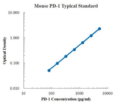
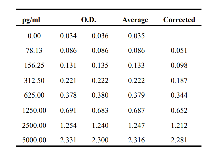
| Sensitivity | 1.61 pg/mL |
| Array Range | 78.13 pg/mL – 5000 pg/mL |
| Assay Time | 3.5 h |
| Recovery | 88% – 126% |
| Average Recovery | 103% |
| Intra Precision | 1.9% – 2.3% |
| Inter Precision | 2.6% – 3.0% |
| Plate | Detachable 96-well plate |
| Storage | If the reagent kit is unopened, it should be stored at 4℃. However, if it has been opened, the standard solution should be stored at -20℃, while the other components should be stored at 4℃. |
| Delivery | 4℃ blue ice transportation |
| Components | 96-well polystyrene enzyme-linked immunosorbent assay (ELISA) plate coated with anti-PD-1 monoclonal antibody Mouse PD-1 freeze-dried standard PD-1 detect Antibody Standard Diluent Assay Buffer(10×) Substrate TMB Stop Solution Washing Buffer(20×) Sealing Film |
| Assay Principle | This kit utilizes the double antibody sandwich enzyme-linked immunosorbent assay (ELISA) detection technique.Specific anti-mouse PD-1 antibodies are precoated on a high-affinity ELISA plate. Standard samples, test samples, and biotinylated detection antibodies are added to the wells of the ELISA plate. After incubation, PD-1 present in the samples binds to the solid-phase antibodies and the detection antibodies. After washing to remove unbound substances, streptavidin-HRP labeled with horseradish peroxidase is added. After washing, a colorimetric substrate, TMB, is added and the plate is incubated in the dark for color development. The intensity of the color reaction is directly proportional to the concentration of PD-1 in the samples.A stop solution is added to terminate the reaction, and the absorbance value is measured at a wavelength of 450 nm (with a reference wavelength range of 570-630 nm). |
Related Targets
PDCD1
PDCD1 Target Infomation Overview
- Target Symbol: PDCD1, programmed cell death 1
- Gene Groups: CD molecules; V-set domain containing
- Alias: CD279; PD1; hSLE1; PD-1
- Previous Names: SLEB2
- Alias Names: systemic lupus erythematosus susceptibility 2
PDCD1, programmed cell death 1 Target Infomation by Species
- Human
- Mouse
- Rat
Human PDCD1 Target Information
- Target Symbol: PDCD1, programmed cell death 1
- Alias:
- CD279
- hPD-1
- hPD-l
- hSLE1
- PD-1
- PD1
- programmed cell death protein 1
- SLEB2
- systemic lupus erythematosus susceptibility 2
- NCBI_Gene: 5133
- UniProtKB: Q15116
Human PDCD1 Predicted Functions
Involved in positive regulation of T cell apoptotic process. Predicted to be located in plasma membrane. Predicted to be active in external side of plasma membrane. Implicated in autoimmune disease (multiple); hepatitis B; hepatitis C; hepatocellular carcinoma; and lupus nephritis. Biomarker of several diseases, including Cryptococcal meningitis; anogenital venereal wart; cervix uteri carcinoma in situ; liver disease (multiple); and tuberculosis (multiple).
Mouse Pdcd1 Target Information
- Target Symbol: Pdcd1, programmed cell death 1
- Alias:
- PD-1
- Pdc1
- programmed cell death
- NCBI_Gene: 18566
Mouse Pdcd1 Predicted Functions
Involved in negative regulation of immune response. Acts upstream of or within negative regulation of apoptotic process; negative regulation of tolerance induction; and positive regulation of apoptotic process. Located in external side of plasma membrane. Is expressed in retina. Used to study dilated cardiomyopathy and systemic lupus erythematosus. Human ortholog(s) of this gene implicated in autoimmune disease (multiple); hepatitis B; hepatitis C; hepatocellular carcinoma; and lupus nephritis. Orthologous to human PDCD1 (programmed cell death 1).
Rat Pdcd1 Target Information
- Target Symbol: Pdcd1, programmed cell death 1
- Alias:
- LOC102550808
- LOC301626
- programmed cell death protein 1
- programmed cell death protein 1-like
- NCBI_Gene: 301626
- UniProtKB: D3ZIN8
Rat Pdcd1 Predicted Functions
Predicted to be involved in negative regulation of immune response and positive regulation of T cell apoptotic process. Predicted to act upstream of or within negative regulation of apoptotic process; negative regulation of tolerance induction; and positive regulation of apoptotic process. Located in plasma membrane. Biomarker of anti-basement membrane glomerulonephritis; periodontitis; and pre-eclampsia. Human ortholog(s) of this gene implicated in autoimmune disease (multiple); hepatitis B; hepatitis C; hepatocellular carcinoma; and lupus nephritis. Orthologous to human PDCD1 (programmed cell death 1).
Products with the Same Target - PDCD1

Competitive ELISA: Principles, Methods, and Key Differences
Competitive ELISA is a widely used immunoassay technique for quantifying target analytes in samples by measuring their interference with a known signal. This method is based on the principle of limited antibodies, where the target analyte competes with a labeled antigen for antibody binding. Competitive ELISA is commonly used for detecting small molecules with fewer epitopes and is available in direct and indirect formats. This article explains the fundamental principles, experimental workflow, and key differences between direct and indirect competitive ELISA, helping researchers choose the optimal method for their applications.

ELISA (Enzyme-Linked Immunosorbent Assay): Principle, Types, and Step-by-Step Protocol
Learn about ELISA (Enzyme-Linked Immunosorbent Assay), a highly sensitive immunoassay technique used in medical diagnostics and biopharmaceuticals. This guide covers its principles, common types (sandwich, direct, indirect, competitive), detailed protocol, and key considerations for accurate results.

ELISA Blood Samples: Serum vs Plasma - Key Differences, Preparation & Hemolysis Effects
Learn whether to use serum or plasma for ELISA testing, including preparation protocols, key differences (fibrinogen, clotting factors), and how hemolysis impacts results. Essential guide for accurate immunoassays.
Related products
-

-
- EK1218
- ELISA Kit
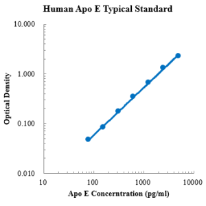
Human Apolipoprotein E/Apo E ELISA Kit
- $299.00 – $419.00
- Select options This product has multiple variants. The options may be chosen on the product page
-
-
-
- EK1188
- ELISA Kit
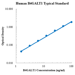
Human B4GALT1/GGTB2 ELISA Kit
- $299.00 – $419.00
- Select options This product has multiple variants. The options may be chosen on the product page
-
-
-
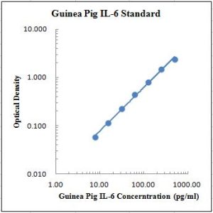
- EK406
- ELISA Kit
Guinea Pig IL-6 ELISA Kit
- $299.00 – $419.00
- Select options This product has multiple variants. The options may be chosen on the product page
-
-
-
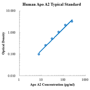
- EK1258
- ELISA Kit
Human Apolipoprotein AII/Apo A2 ELISA Kit
- $299.00 – $419.00
- Select options This product has multiple variants. The options may be chosen on the product page
-
-
-
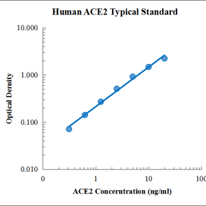
- EK1391
- ELISA Kit
Human ACE2 ELISA Kit
- $299.00 – $419.00
- Select options This product has multiple variants. The options may be chosen on the product page
-
-
-
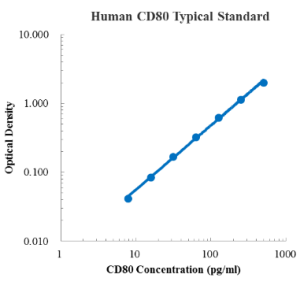
- EK1272
- ELISA Kit
Human B7-1/CD80 ELISA Kit
- $299.00 – $419.00
- Select options This product has multiple variants. The options may be chosen on the product page
-
Reviews
There are no reviews yet.